Which X-ray Would Be Used to Visualize the Gallbladder
Because only 10 of all gallstones are calcified this imaging study has limited usefulness. Contrast dye is then used to visualize these ducts on an X-ray.
Gallstones Radiology Reference Article Radiopaedia Org
An ultrasound allows doctors to view images of the organs and soft tissues inside your body.

. An X-ray of the gallbladder can be viewed through an abdominal X-ray which sometimes requires the patient to swallow a dye or have it injected into the body in order to create a clearer image of the gallbladder. It can sometimes detect kidney stones an obstruction blockage a perforation hole in the intestines or an abdominal mass such as a tumor. Which of the following endoscopic exams would be used to view inside a section of the colon.
An x-ray uses electromagnetic radiation to create black-and-white images of the inside of your body. Some X-ray types require that a patient swallow a dye or have dye injected into the body so the X-ray can capture a. A computed tomography CT scan constructs detailed X-ray.
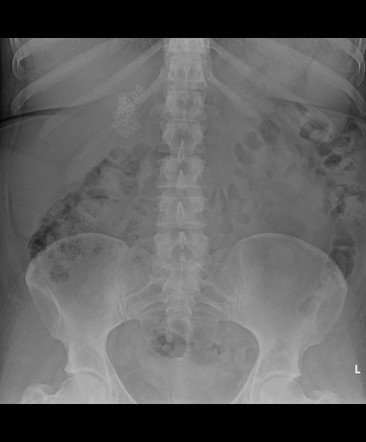
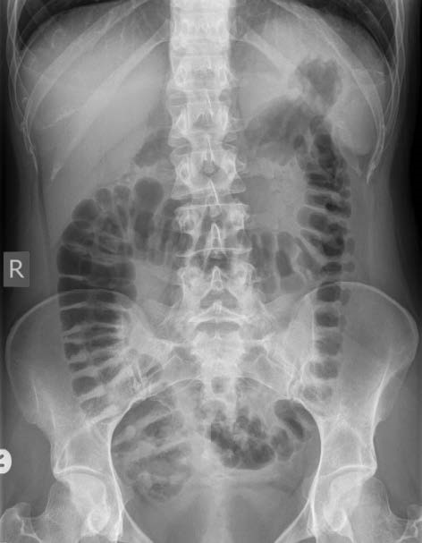
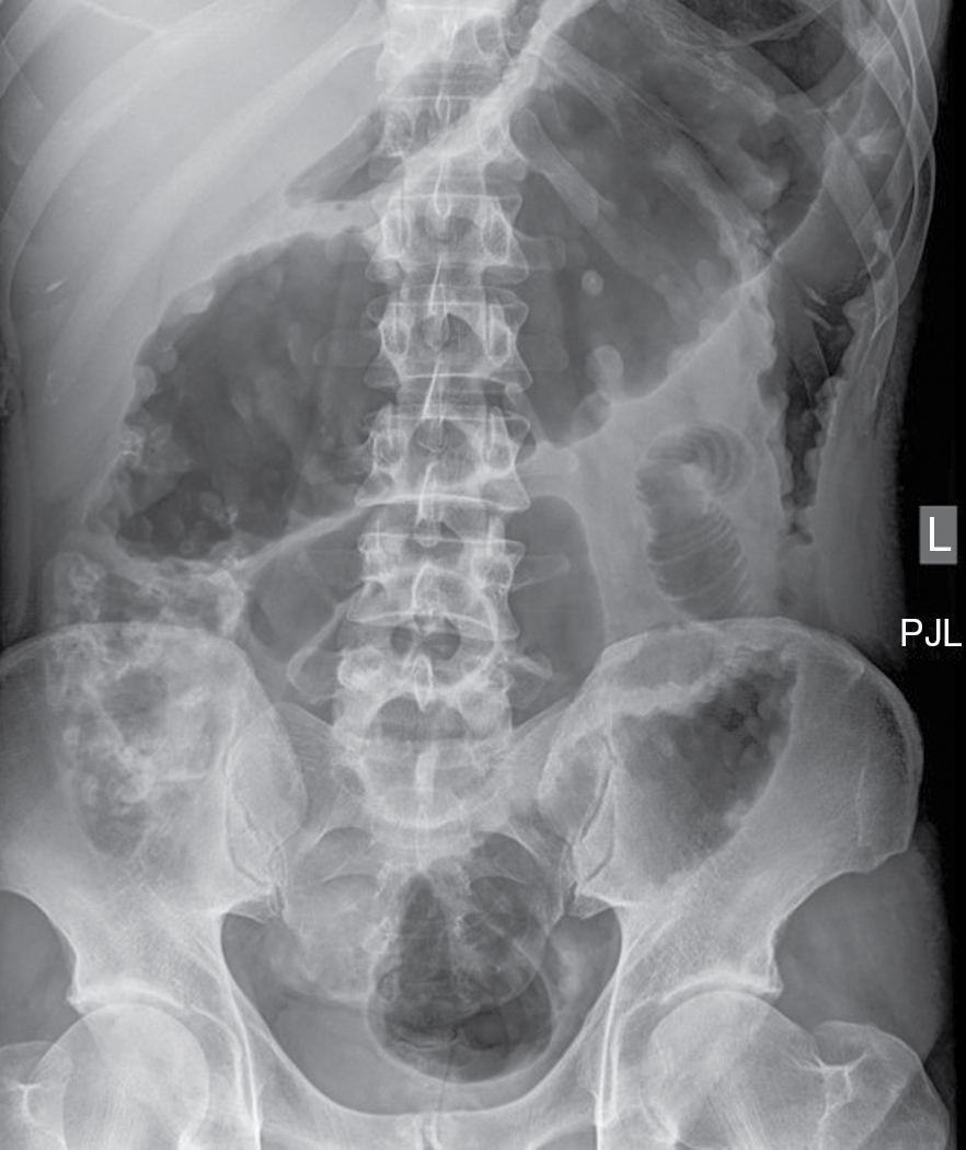
The following day at the hospital the radiologist examines the gallbladder with a fluoroscope a special x ray that projects the image onto a video monitor. X-Ray of Abdomen X-ray of the abdomen usually done for other purposes can show big calcified gallstones in the gallbladder. The image also can reveal small metal objects such as coins that might have been swallowed.
It should empty freely with no obstruction after the PFM post-fatty meal. There are several diagnostic tests used make the determination of gallbladder disease. However CT scans also can miss gallstones that you may have.
In some cases you may need to swallow a substance. Ultrasonography Ultrasonography Ultrasonography uses high-frequency sound ultrasound waves to produce images of internal organs and other tissues. In gallbladder infection the air in the gallbladder can be sometimes seen.
Other related procedures that may be used to diagnose problems of the gallbladder include abdominal X-rays computed tomography CT scan of the liver and biliary tract abdominal ultrasound cholecystography or endoscopic retrograde cholangiopancreatography ERCP. Unlike a generalized abdominal ultrasound a gallbladder ultrasound often referred to as right upper quadrant ultrasound is a more specialized procedure to test the gallbladder and ducts attached to it and the patient undergoing the procedure may need to order further tests later. X-ray image of the gallbladder.
It is also used to look for bile duct blockage or leak. Porcelain gallbladders can also be seen in plain x-rays. CT scans use a combination of x-rays and computer technology to create images of your pancreas gallbladder and bile ducts.
The procedure for getting an x-ray can vary. Gallbladder radionuclide scan Gallbladder radionuclide scan is a test that uses radioactive material to check gallbladder function. Your health provider may order an x-ray if they suspect gas buildup calcified gallstones or porcelain gallbladder concerns.
Using sound waves an ultrasound provides a real-time picture of your. Having a view of your bile duct during surgery. CT scans can show gallstones or complications such as infection and blockage of the gallbladder or bile ducts.
Which medication is used to treat constipations. You could need other tests along with an x-ray. Dye is administered intravenously to the patient allowing for X-ray visualization of the gallbladder and bile ducts.
Its usually done during surgery to remove your gall bladder. Chest X-ray for Gallbladder Cancer About Clinical Trials Our Clinics Overview Symptoms Causes Diagnosis Physical exam history Ultrasound CT scan Blood chemistry studies Liver function tests Carcinoembryonic antigen assay CA 19-9 assay Chest X-ray MRI Endoscopic retrograde cholangiopancreatography Biopsy Laparoscopy. Why is it done.
Reasons for a Gallbladder X-Ray. How the Test is Performed The health care provider will inject a radioactive chemical called a gamma-emitting tracer into a vein. A simple abdominal x-ray can be used to identify calcified gallstones.
An abdominal X-ray can spot gas and some types of gallstones containing calcium. An intraoperative cholangiogram IOC is an X-ray of your bile ducts. The use of ultrasound testing which uses sound waves to image and make a picture of the intra-abdominal organs including the gallbladder.
An abdominal X-ray may help to find the cause of abdominal pain or vomiting. Sometimes patients are then asked to drink a highfat formula that will cause the gallbladder to contract and release bile. The following day at the hospital the radiologist examines the gallbladder with a fluoroscope a special x ray that projects the image onto a video monitor.
Sometimes patients are then asked to drink a highfat formula that will cause the gallbladder to contract and release bile. What is a gallbladder ultrasound. A physician may order an X-ray to check for certain cancers in different parts of the body by.
X rays will then be taken at various intervals. Results A normal OCG will show a normal gallbladder. The x-ray technologist should be informed since the gallbladder might not be well visualized.
Transabdominal ultrasonography is better for detecting. Magnetic resonance imaging MRI. A device called a transducer converts electrical current into sound waves.
X rays will then be taken at various intervals. X-rays are used for a multitude of reasons. Read more uses sound waves to provide images of the liver gallbladder and bile ducts.
The gallbladder should visualize and be free of any solid structures such as stones polyps or tumors. Which x-ray would be used to visualize the gallbladder.

Abdominal X Ray Showing Two Plastic Stents Double Pigtail In The Gall Download Scientific Diagram

Approach To The Abdominal X Ray Axr Undergraduate Diagnostic Imaging Fundamentals
Approach To The Abdominal X Ray Axr Undergraduate Diagnostic Imaging Fundamentals
Abdominal Radiography Radiology Key
0 Response to "Which X-ray Would Be Used to Visualize the Gallbladder"
Post a Comment